Real-Time Ultrasound Biofeedback
Real-Time Ultrasound Biofeedback is a non-invasive imaging technique that uses diagnostic ultrasound to visualize muscles and organs in motion while you perform specific movements or breathing tasks. In physical therapy, it serves as a visual biofeedback tool, helping you see deep muscles such as the transversus abdominis, multifidus, pelvic floor, and diaphragm as they contract and relax, improving precision and control of these stabilizing systems.
Mechanism of Action:
Real-Time Ultrasound Biofeedback promotes:
Visualization of deep stabilizing muscles (e.g., transversus abdominis, multifidus, pelvic floor, diaphragm) during contraction and relaxation
More accurate and selective activation of targeted muscles through real-time visual feedback
Improved motor learning, timing, and coordination of core and breathing muscles
Awareness of compensatory patterns, such as overuse of superficial muscles or breath-holding strategies
Refinement of breathing mechanics and pressure management through observation of diaphragm and abdominal wall motion
Clinical Applications:
Real-Time Ultrasound Biofeedback is used to support:
Chronic or recurrent low back pain through retraining of transversus abdominis and lumbar multifidus activation and endurance
Pelvic floor rehabilitation and continence training, including coordination with the diaphragm and abdominal wall
Postpartum and post-surgical core rehabilitation (e.g., after abdominal or pelvic procedures) where visualizing deep support muscles improves safety and effectiveness
Breathing pattern disorders and diaphragm retraining in patients with neck, thoracic, or stress-related dysfunction.
Optimization of trunk stability and lumbopelvic control in dancers, athletes, and performers who require fine motor control under load.
Education and neuromuscular re-education in complex pain presentations where improving body awareness and self-efficacy is essential.
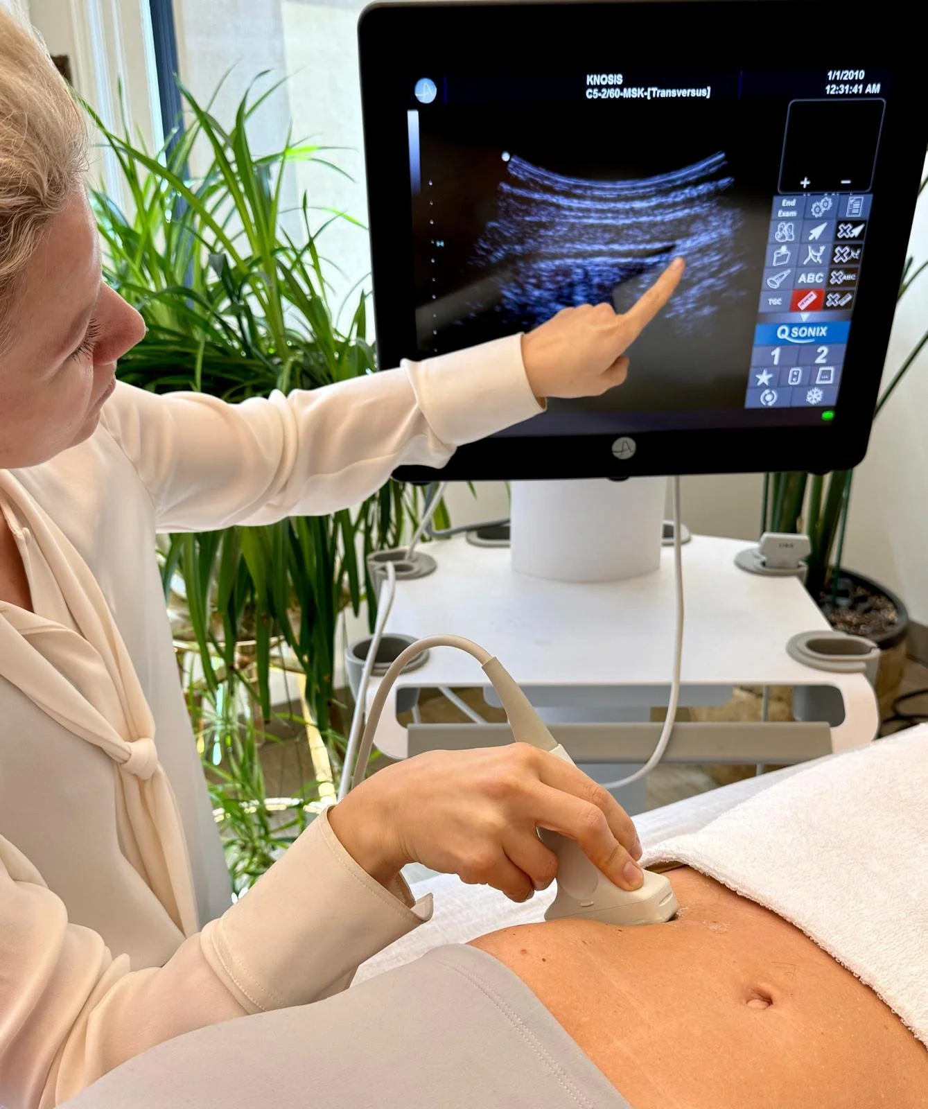
Procedure
During a session, a small ultrasound probe is placed over the region of interest (for example, the lower abdomen, lumbar spine, pelvic floor region, or lower ribs). As you perform gentle activation tasks—such as drawing in the lower abdomen, contracting the pelvic floor, or taking diaphragmatic breaths—the ultrasound image shows your deep muscles moving in real time on a monitor. The therapist coaches you to adjust the quality, timing, and intensity of your contractions based on what you see, helping you learn how correct activation feels and how to reproduce it during everyday movement and exercise.
Scientific Basis
Rehabilitative ultrasound imaging (RUSI) is recognized as a valid and reliable method for assessing the thickness and contraction of deep trunk and pelvic muscles, including the transversus abdominis, lumbar multifidus, and pelvic floor.
Randomized and controlled studies show that adding real-time ultrasound visual biofeedback improves the ability to activate and retain activation of muscles such as the multifidus and deep abdominals compared with verbal instruction alone.
Research in pelvic health and breathing science further supports ultrasound as an effective tool to assess and retrain pelvic floor and diaphragm motion, enhancing patient understanding and engagement in rehabilitation.
At KNOSIS, Real-Time Ultrasound Biofeedback is integrated within comprehensive treatment plans to refine core, pelvic floor, and breathing strategies—linking deep stability with posture, movement, and nervous system regulation to support durable, functional change.